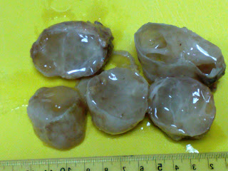
40 years old, Bangali Female patient, has many discharging breast sinuses. Received breast tissue shows multiple cutaneous sinuses, and cut section shows many irregular brownish cavitations oozing yellowish material. Microscopically shows non-caseating epithelioid granulomas with lympho-plasmacytic infiltrate and focal central neutrophilic collection as well as scattered multinucleated giant cells. ZN stain was negative for AFB. PAS stain shows positive needle-shaped bodies within the center of some granulomas ; suggestive of Actinomycosis.
Tuesday, November 29, 2011
Monday, November 28, 2011
Ovarian Mesonephroid Adenocarcinoma + Bilateral Ovarian & Cervical Endometriosis
Philipino lady, 46 YO, C/O Severe lower abdominal pain and intermittent bleeding. Sonography shows Lt. ovarian cyst 7 cm in diameter (not shown in photos). Frozen section shows a malignant growth. Total hysterectomy with bilateral salpingo-opherctomy was done. Histopathologic examination shows invasion of Lt. ovary by malignant mixed papillary and tubular growth with clear cells and focal hobe-nail pattern; bilateral ovarian endometriosis and cervical endometriosis.
Wednesday, November 16, 2011
MULTIPLE INTRAMUSCULAR MYXOMAS
Sudanese female, 60 years old, with multiple Rt. thigh swellings, appeared on ultrasonography as multiple thigh lipomas. Gross: 3 grayish well defined partially cystic masses, the largest 9.5X5 cm and the smallest 6X3 cm. Cutsection shows mucoid content with firm solid grayish parts. Microscopic: hypocellular pattern, with bluish mucoid background, no mitoses, few coarse BVs, peripheral muscle and fat tissue.(No lipoblasts)
Saturday, October 15, 2011
Prostatic SEMINOMA (case report)
72 years old Nigerian male diagnosed clinically and radiologically to have prostatic tumor. PSA, AFP and CEA were within normal serological values. TURP was done and the diagnosis was seminoma. This was followed by bilateral orchidectomy. Both testes were negative for malignancy, however, they were atrophic and have scattered bilharzia ova. So, it is primary prostatic seminoma (6th case in literature).Tissue markers are currently done for confirmation.
Tuesday, April 19, 2011
Cervical lymphadenopathy
Male patient, 55 YO, with moderately enlarged cervical lymph nodes, firm> No hepatic or splenic enlargement by sonar and showing recent gradual weight loss.
Received multiple LNs; the largest 2.5 cm in diameter with grayish cut surface.
Microscopically; loss of architecture, infiltration by groups of large cells with vesicular nuclei and prominent nucleoli, scattered mitoses, irregular fibrosis and plasma-lymphocytic infiltration. (DD;sarcomatoid variant of ALCL; follicular dendretic lymphoma, inflammatory pseudotumor, ....OTHERs. Plz, what are ur suggestions and recommended markers.Thanks.
Subscribe to:
Comments (Atom)


